Learning Center: Access information in greater detail.
Tumors in the Nose and Sinuses
Use the search tool, scroll down for your topic of interest, or use the index at the bottom of this page.
sinus anatomy
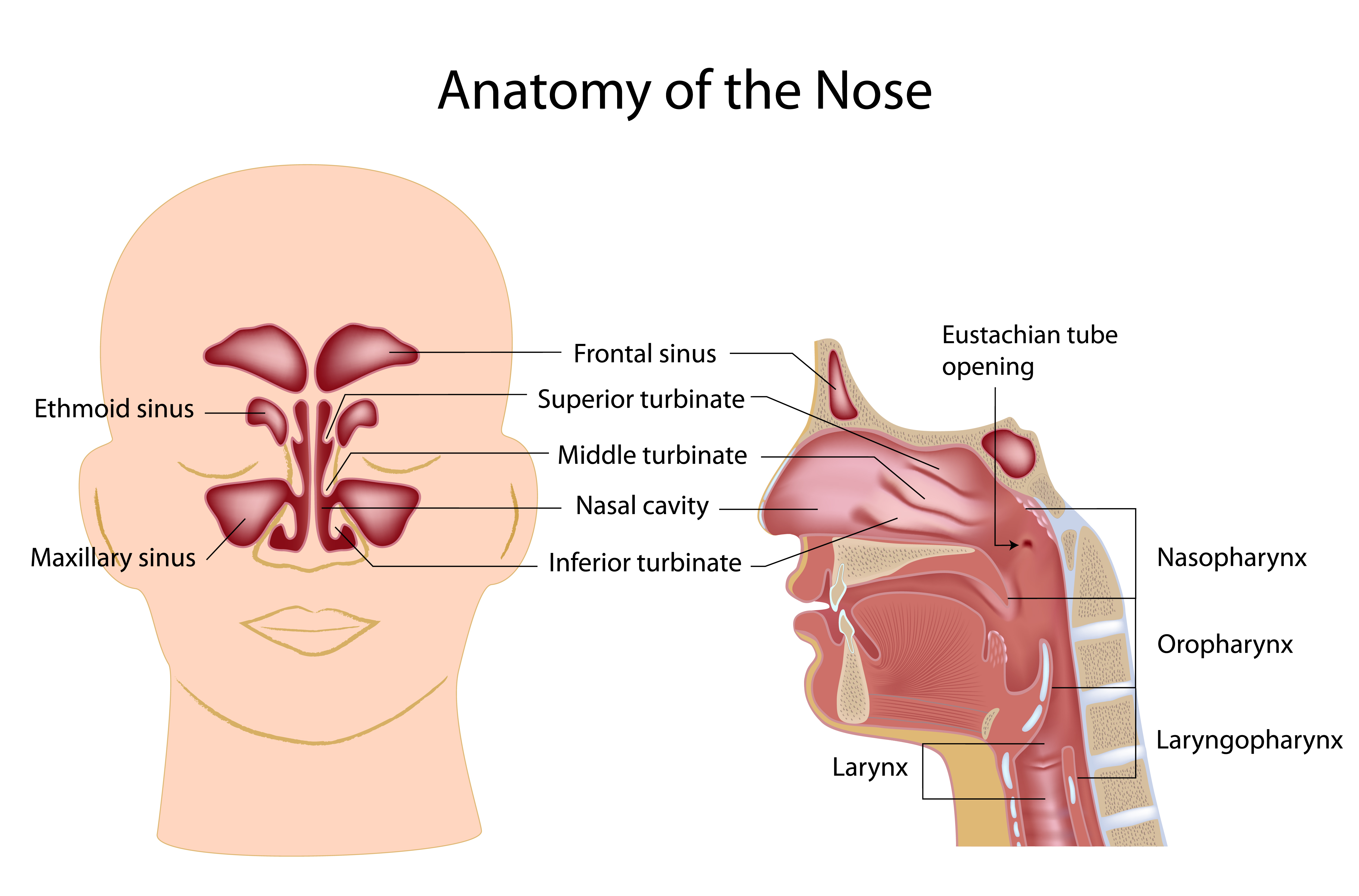
There are four pairs of sinuses surrounding the nasal cavity:
The maxillary sinuses, the largest of the paranasal sinuses, are under the eyes, in the maxillary bones.
The frontal sinuses, superior to the eyes, in the frontal bone, which forms the hard part of the forehead.
The ethmoidal sinuses, which are formed from several discrete air cells within the ethmoid bone between the nose and the eyes.
The sphenoidal sinuses are located behind (posterior to) the ethmoid sinuses, in the sphenoid bone.
The paranasal air sinuses are lined with respiratory epithelium (ciliated pseudostratified columnar epithelium), which has microscopic cilia, which are fingerlike projections that move in a coordinated fashion to sweep mucus over the surface toward the nasal cavity.
The nasal cavity is the air-filled space above and behind the nose in the middle of the face. The nasal septum divides the cavity into two cavities. Each nasal cavity is the continuation of one of the two nostrils. The nasal cavity is the uppermost part of the respiratory system and provides the nasal passage for inhaled air from the nostrils to the nasopharynx and rest of the respiratory tract. The nasal cavity is divided in two by the vertical nasal septum. On the side of each nasal cavity are three horizontal outgrowths called turbinates, the inferior, middle, and superior turbinates. These turbinates warm and humidify airflow before it reaches the throat and lungs. The turbinates also disrupt the airflow, directing some air toward the olfactory epithelium between the middle turbinates and the septum.
Cancers of the paranasal sinuses comprise approximately 0.2% of all malignancies. About 80% of these malignancies arise in the maxillary sinus. Men are much more often affected than women. They most often occur in the age group between 40 and 70 years. Carcinomas are more frequent than melanomas and sarcomas. Metastases to the sinuses from a cancer starting elsewhere are rare.
evaluation of sinonasal tumors
Because tumors within the nose or sinuses are not easily seen, they are often identified after significant growth has occurred. The symptoms that typically indicate a problem include nosebleeds, nasal airway blockage (especially if one sided and progressively worse), numbness on the cheek skin, double vision, excessive tearing from one eye (epiphora), difficulty opening the mouth widely (trismus), teeth not fitting together well (malocclusion), or development of a neck mass from spread to a neck lymph node. Physical exam, fiberoptic endoscopy, and imaging (CT, MRI, and/or PET-CT scan), and biopsy are commonly performed for evaluation, before a treatment strategy can be outlined.
types of sinonasal tumors
Benign sinonasal tumors include papillomas (inverting, fungiform, and cylindrical cell subtypes), osteomas (boney growths). Malignant (cancerous) sinonasal tumors include squamous cell carcinoma, adenocarcinoma, adenoid cystic carcinoma, and less commonly melanoma, sarcoma, or neuroendocrine cancers.
Treatment for SINONASAL TUMORS
Benign tumors, such as inverted papilloma, are usually treated surgically, and with an endoscopic approach if the tumor location and extent is favorable. A benign sinus osteoma is typically not treated unless it is blocking the outflow of a sinus. Malignant tumors, when treatment is intended for cure, are often treated with surgery followed by radiation therapy or chemotherapy plus radiation therapy. Depending on the location of the tumor, the surgical defect may require tissue reconstruction or use of a special denture called an obturator.
additional links
Learning Center Main Index:
Throat:
swallowing, tonsils and adenoids, obstructive sleep apnea, voice
Aesthetics:
skin regimen, injectables {neuromodulators (e,g. Botox), hyaluronic acid fillers (e.g., Juvederm), and others}, rhinoplasty, facelift, neck lift, and brow lift, blepharoplasty (eyelid surgery), skin resurfacing, scar treatment
Tumors (benign and malignant/cancerous):
general tumor information, thyroid, parathyroid, skin, neck, oropharynx, larynx (voice box), salivary gland, nose and sinus, oral cavity (mouth and lips), nasopharynx, hypopharynx, radiation therapy, chemotherapy and immunotherapy, gastric feeding tube
Nose and Sinus:
rhinoplasty (functional and cosmetic), sinusitis, breathing