Diagnostics: Pathology (biopsy)

Tissue that is removed may be sent to pathology for analysis. The surgical pathologist will examine the tissues and provide a report summarizing the data gleaned, including the diagnosis of the disease process, if present. Additional information relevant to the disease process is provided as well.
Some of the more common methods of pathological analysis are described below.

Squamous cell carcinoma processed with standard histopathology technique viewed in a low power (zoomed out) microscope view.
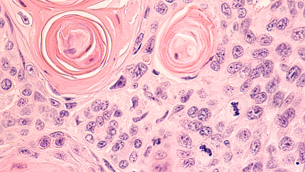
Squamous cell carcinoma processed with standard histopathology technique viewed in a high power (zoomed in) microscope view.
Histopathology is the examination of a biopsy or surgical specimen by a pathologist, after the specimen has been processed and histological sections have been placed onto glass slides. During a surgical operation, a tissue specimen may be analyzed with a modified histopathology technique called frozen section processing, which provides results within about 30 minutes of submission of the specimen. Frozen section technique is used when the results would be actionable to the surgeon during the operation. More commonly, when not dealing with a situation requiring quick results during an ongoing operation, permanent section histopathology is performed. Permanent section pathologic diagnosis is more accurate than with frozen sections, but it takes days or weeks typically to have the final results. Specimens sent for frozen section pathology are usually divided and one portion is processed for permanent section analysis as a more definitive, albeit slower, analysis of the same tissue. Permanent section histopathology allows the pathologist to perform special stains and immunohistochemistry to evaluate the molecular properties of the tissue, aiding in diagnosis and characterization of the tissue.

Microscopic view of a fine needle aspiration specimen of a lymph node containing squamous cell carcinoma. Since the squamous type of cell does not normally exist in a lymph node, its presence in a lymph node indicates that this type of cancer has spread from somewhere else to this lymph node.
Cytopathology examines free cells or tissue micro-fragments of cells collected from a fine needle aspiration biopsy. Analysis of a cytopathology specimen involves looking at a small number of cells that have been separated from each other, thus depriving the pathologist of clues such as the arrangement or architecture of cells that a larger specimen processed by histopathology would provide.
Flow cytometry is a technique used to analyze a population of cells obtained in a biopsy. This technique is primarily used in medicine to determine if a specific category of cancer, lymphoma, is present. A specimen for flow cytometry is sent to the laboratory in a container free of preservative and analyzed expediently to maximize the portion of cells that are alive and therefore useful.

This age