Diagnostics: Ultrasound
What is an ultrasound imaging study?
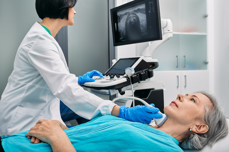
An ultrasound imaging study, also known as ultrasonography or sonography, is a non-invasive medical procedure that utilizes high-frequency sound waves to produce visual images of the body's internal structures. It is commonly used to examine various organs, tissues, and systems, including the thyroid, parathyroid, neck, abdomen, pelvis, heart, blood vessels, and reproductive organs. During the procedure, a small handheld device called a transducer is gently moved over the skin, transmitting sound waves into the body. These waves bounce off the structures inside and create echoes, which are then captured by the transducer and converted into real-time images on a monitor. Ultrasound imaging provides valuable diagnostic information, and is sometimes the most informative type of imaging for a given condition. Ultrasound is limited by the inability of the sound waves to penetrate bone, thus making imaging of structures behind bones very limited. The procedure is safe, painless, and does not involve the use of ionizing radiation, making it a widely used and valuable tool in medical practice.
Ultrasonography is also used frequently to assist in the accurate placement of a needle for biopsy purposes, as in an ultrasound guided fine needle aspiration biopsy and an ultrasound guided core needle biopsy.

HOW TO GET THE MOST FROM YOUR APPOINTMENT
Appointment time is valuable. Below are some suggestions to make the most of your appointment. This preparation will help you and your doctor maximize efficiency and accuracy, freeing up time for questions and answers.
• Click here to prepare for your neck mass/swelling/lump appointment.

This page