Treatments: Skin Cancer
The vast majority of skin cancer is treated with surgery alone. More advanced cancers, those that are large or have spread, however are often treated in multiple ways, such as surgery and radiation or medicine. On occasion, if a patient is not a good surgical candidate or if surgery would not be helpful, non-surgical treatment is used.

Most skin cancers are treated with high rates of cure with surgical excision. There are various techniques for doing this, including local excision and Mohs surgery. With local excision, the cancer plus an additional amount of normal tissue surrounding it. A pathologist then uses a microscope to look at the specimen provided to see whether the cancer is fully contained within a rim of normal tissue. This can be performed in real-time by a technique called frozen section. The results are relayed back to the surgeon, and if cancer extends all the way to the edge of the excised tissue or if it is too close to the edge, additional tissue may be removed all in the same operation. Mohs surgery is very similar to this. In Mohs micrographic surgery, the dermatologist acts as both surgeon and pathologist, and the orientation of the specimen on the microscope slide is horizontal instead of vertical.

adjacent tissue transfer (local flaps)
If the area missing skin after removal of a skin cancer is too large to close well by simply stitching the edges together, techniques of borrowing nearby tissue making use of its stretch, skin characteristics, and blood supply are used in local tissue transfer (also called local flaps).
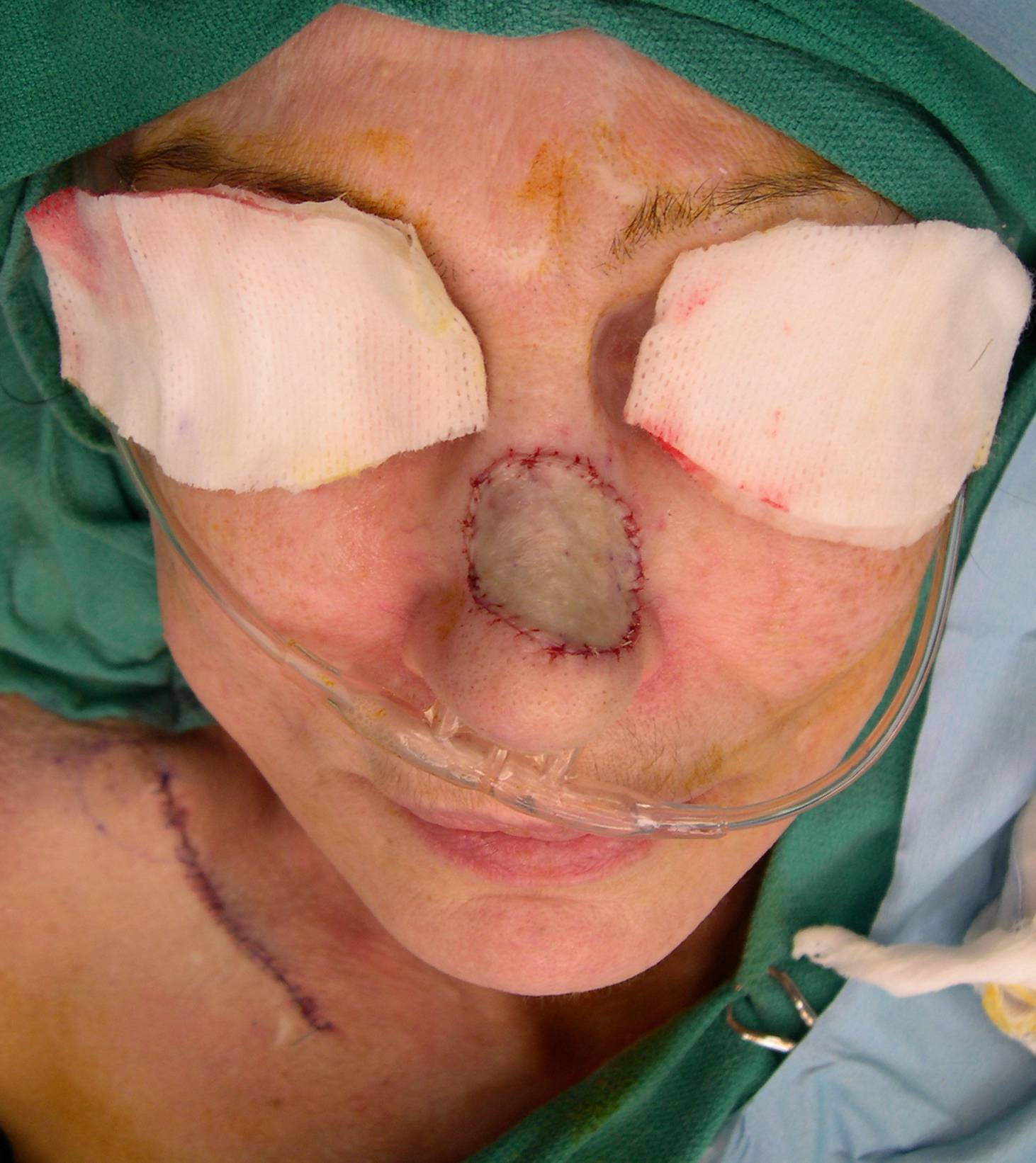
Skin grafts
Another method of reconstructing a skin defect is to use a skin graft, which may be the top layers of skin (a partial thickness skin graft) or the full thickness of skin (a full thickness skin graft). This skin graft is secured under a dressing at the skin cancer site and allowed to heal. The area where the skin graft was taken from is also prepared for healing. At about a week after the procedure, the dressings and sutures are removed. As the skin heals, the color and texture improve.
Lymph node removal (lymphadenectomy or “neck dissection”)
If one or more lymph nodes in the neck have cancer or are at high risk of having cancer spread to them, those lymph nodes typically need to be treated. One method of dealing with these neck lymph nodes is to remove them surgically. This is an effective treatment as well as very informative from a diagnostic standpoint, as the number of lymph nodes with cancer and any adverse features present help to tailor any additional treatments.

Sentinel lymph node biopsy
The sentinel lymph node is the hypothetical first lymph node or group of nodes draining (filtering fluid from) a cancer. It is postulated that the sentinel lymph node is the most likely lymph node to be involved by a cancer if spread has occurred. Therefore, removing and testing a sentinel lymph node can indicate whether spread has occurred, even if too small to be felt on exam or seen on x-rays or other imaging. The sentinel node biopsy is the identification, removal and analysis of the sentinel lymph node (or sentinel lymph nodes if a few good candidates are apparent) of a particular tumor.

External beam radiation (radiation therapy)
Radiation therapy is an effective type of treatment for many skin cancers, especially small ones. The cost of radiation is greater than that of surgery and the time involved is greater as well. In addition, radiation affects the treated tissues such that healing in an area previously radiated is compromised and repeating radiation in a given area has significantly higher risks. For these reasons, radiation is considered by many to be a second choice for the initial treatment of most skin cancers.
Radiation therapy may be used after surgery in situations where the likelihood of cure would otherwise be too low. For example, if surgical results show that a skin cancer is an aggressive type (such as Merkel cell carcinoma), or if there is a reasonable suspicion that some cancer is left behind (with tumor tracking along nerves or the excision not getting around the tumor completely), radiation therapy may be advised. Radiation could be applied to the site of the original tumor as well as to areas where the tumor may have spread, such as to the lymph nodes in the area.

Medical therapy (chemotherapy and immunologic therapy)
For some skin cancers that exist only on the top layer of the skin (epidermis), medicated creams may provide reasonable cure rates. For example, for basal cell carcinomas that involve only the epidermis, 5-FU (fluorouracil) can be used with a 90% cure rate and imiquimod may be used to treat thin basal cell carcinomas with a 70% cure rate. These cure rates do not apply to basal cell carcinomas that extend deeper than the top layer of skin, and the only way to be sure of the depth of a basal cell carcinoma is by biopsy or full removal of the cancer. Thus, these methods are less than ideal, but have a role when other methods (surgery or radiation) cannot be used.

The best treatment: Prevention or early detection of skin cancer
Even after skin cancer has been identified and treated, you have the ability to prevent additional skin cancers from forming by diligently protecting your skin from ultraviolet light. After one skin cancer is identified, an individual has a 50% chance of having another skin cancer within three years. Early detection makes treatment easier and more successful.

This page