Conditions: Lymph Node
Lymph nodes are part of the immune system protecting us from infection. When activated to fight infection, a lymph node becomes larger and tender. Some other conditions also make a lymph node larger, including cancer. Generally, if an enlarged lymph node is tender and has been present for less than seven weeks, it is very likely to be reacting appropriately to an infection. Lymph nodes that are not tender, grow progressively and unrelentingly for more than seven weeks, and are not tender may benefit from medical evaluation.
enlarged Lymph node

The neck contains many lymph nodes, and these are usually too small to be seen or felt. Enlargement of one or more neck lymph nodes can occur as a result of inflammation (such as with infection) or growth of a tumor within lymph node(s).
Most often, enlarged neck lymph nodes arise from nearby infection, such as tonsillitis (infection of the tonsils) for example. More concerning is persistent enlargement (and especially unrelenting growth) of a lymph node because this may represent a situation in which cancer is in this lymph node, either having spread from somewhere else (as in squamous cell carcinoma, for example) or starting within the lymph node (as in lymphoma). Because the vast majority of swollen lymph nodes are enlarged because of infection, not every noticeable lymph node needs to be evaluated. When a lymph node has been present for several weeks (roughly seven or longer), when the affected individual has risk factors (such as a history of smoking or having had cancer previously, or with other symptoms (trouble swallowing, a wound in the mouth, or a non-healing skin wound, for example), evaluation by a physician may be warranted.
Lymph nodes may become enlarged in cancerous (malignant) disease. Cancer, such as squamous cell carcinoma from the mouth or throat, can spread to one or more lymph nodes and grow in that structure. Alternatively, persistently enlarged lymph nodes may represent a cancer arising in the lymph node itself, as in lymphoma (both Hodgkin's and non-Hodgkin's) or lymphocytic leukemia, Lymph node enlargement that lasts less than seven weeks or more than one year with no progressive size increase has a low likelihood of being neoplastic.
It is important to note that a lymph node enlargement may be due to 1) an appropriate reaction of the lymph node as it responds to nearby infection, 2) cancer, or 3) any of a number of other conditions such as sarcoidosis. Generally, the larger the lymph node, the greater the chance of it harboring cancer. Also, progressive growth of a lymph node is more suspicious for cancer than fluctuation in size without any trend of growth over weeks to months.
Lymph nodes with cancer spread to them (“metastatic lymph nodes”) become enlarged because tumor cells have detached from the primary tumor, moved to a lymph node, and grown in the lymph node. Since cancer generally occurs somewhat more frequently in late middle age people, this kind of lymph node abnormality is more common in older persons. Metastatic lymph nodes tend to feel hard and may be fixed to underlying tissues and are often not tender to the touch. Usually the lymph nodes that directly drain the area of the cancer are affected by the spread. Sometimes metastatic cervical lymph node is detected before the main cancer. In such cases, this discovery leads to a search for the primary malignancy—where the cancer originally arose.
Now, objectively, what size of lymph node is technically enlarged? It depends, but based on what one can feel with the fingers, a lymph node greater than 1 to 1.5 cm is enlarged. This is a fairly imprecise way to assess lymph node size. With medical imaging, such as ultrasound or CT scan, a lymph node size in three dimensions can be assessed. It turns out that the most important dimension for assessing abnormal enlargement is the length of the shortest axis and the roundedness of the three dimensional structure. In other words, a lymph node measuring 1.8 x 0.8 x 0.6 cm would not usually be considered suspicious for cancer but one measuring 1.8 x 1.6 x 1.7 cm would be considered to have a higher risk of cancer because the shortest axis is greater than 1 -1.5 cm. With imaging, additional image-based information beyond size measurements are available and may also help in assessing suspicion for cancer in a lymph node as much or more than the size alone. As only one example, the presence of fat in the center of the lymph node is normal, but may be absent in a lymph node with cancer growth within it.
What is a lymph node?

A lymph node. Lymphatic fluid flows in to the lymph node at the outer surface (cortex) shown by red arrows. The lymphatic fluid flows out at the center portion of the lymph node (hilum) shown by blue arrows. Blood flows in (red vessels) and out (blue vessels) also via the hilum. In the center of the lymph node, lymphatic cells process a sampling of the material (proteins and cells) passing through; If an abnormality is detected, the lymphatic cells divide and the lymph node swells.

This is a mildly enlarged but benign lymph node.

The mildly enlarged but normal lymph node, cut in two halves, demonstrating the central hilum and the outer cortex.
A lymph node is an organ of the lymphatic system, which is part of the immune system. Many lymph nodes exist throughout the body, and are connected by tiny channels or tubes called lymphatic vessels. The lymph nodes are like the beads on a string, where the string is the lymphatic vessel.

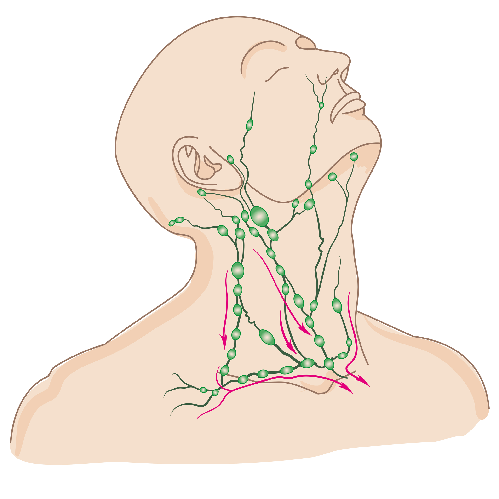
Diagram of the lymph nodes along different lymphatic channels in the torso, arms, and neck. Note the high density of lymph nodes in the neck and arm pits.
A lymph node is a shape roughly similar to a lima bean. It has an outer protective layer, and within it are cells of the immune system. Both lymphatic fluid and blood flow through a lymph node. The immune system cells in the lymph node sample the fluids running through it, looking for a protein or other molecule that should not be present, such as a protein from a virus, bacteria, or cancer. When the cells in a lymph node detect a molecule that should not be present, the immune cells in the node multiply and the lymph node swells, becoming larger and often tender to the touch.
Cancer cells can travel through the lymphatic channels and spread from the original site of a cancer to other parts of the body. Occasionally, a cancer cell flows into a lymph node and, instead of passing right through, it can stay in the lymph node long enough to divide into two cells. Then the two cancer cells divide into four cells, and so on, which can eventually make the lymph node enlarged. This is how cancer spreads to lymph nodes. Often, a lymph node that has become enlarged due to infection is tender (uncomfortable), whereas a lymph node that has become enlarged due to cancer is not uncomfortable.
What is the lymphatic system?

In green is a lymphatic vessel, or tube, that collects fluid, waste products, cellular debris, viruses, bacteria, and potentially even cancer cells from the spaces between surrounding cells.

The lymphatic vessels, shown in green, collect fluid from around the nearby cells, some of which comes from the blood vessels. Another way of looking at it is that the lymphatic vessels are drains for fluid that has leaked into tissue from blood vessels or cells.
The lymphatic system, or lymphoid system, is an organ system that is part of the immune system, and complementary to blood circulation. It consists of a large network of lymphatic vessels, lymph nodes, lymphatic or lymphoid organs, and lymphoid tissues. The vessels carry a fluid called lymph (or “chyle”) back toward the heart, for re-circulation.
Blood flows away from the heart through arteries that branch and become smaller and then feeds into capillaries, where oxygen, nutrients, and some fluid are delivered. Capillaries then join up with small veins that merge with each other, making large veins returning blood back to the heart. Some fluid (plasma) leaks out through capillaries into the tissues. The fluid bathes the tissues as interstitial fluid, and this interstitial fluid mixes with cellular waste products, bacteria, and damaged cells before being drained by lymphatic capillaries. One main function of the lymphatic system is to provide a route for this leaked fluid to return to the bloodstream. If this fluid does not return from tissues to the bloodstream, lymphedema occurs.
The other main function of the lymphatic system is immune defense. As fluid exits the bloodstream, bathes the cells and then collects in lymphatic channels, a sampling of the goings on of the body is accomplished. Lymphatic fluid (also known as “lymph” or “chyle”) contains waste products, cellular debris, proteins, and potentially viruses, bacteria, and/or cancer cells.
As lymph flows back toward the subclavian veins, it passes through lymph nodes, where a high concentration of immune cells called lymphocytes process the contents of the lymph. Lymphocytes recognize proteins that are not part of one’s self, and will become activated and proliferate, orchestrating an effort to send white blood cells to the source of the non-self proteins (such as a bacterial infection) and destroy it. When a lymph node is activated, it becomes larger and tender/uncomfortable, which may bring it to an individual's attention. Other lymphatic organs include the spleen, the thymus, and the tonsils. The tonsils probably complete their function and contribute no more by a very early age, the thymus slows down activity and shrinks considerably by the early teenage years, and the spleen contributes throughout life, especially against certain bacteria noted to be “encapsulated.”

Lymphatic circulation schematized. Fluid that leaks out of the blood circulation (arteries, veins, and capillaries) and fluid that leaks out of cells collects in lymphatic vessels. The smallest lymphatic vessels, called lymphatic capillaries, merge with each other an make larger lymphatic vessels (tubes or channels). Lymph nodes are like filters along the pathway of lymphatic vessels. Eventually, the lymphatic fluid returns to mix in with the venous circulation of blood. Not shown in this diagram is the contribution of long-chain fatty acids absorbed from the diet by the gut into the lymphatic circulation..

Anatomic depiction of the lymphatic system, the collection of lymph from lymphatic capillaries and recirculation into the bloodstream at the subclavian vein via the thoracic duct. Not shown here is the gut and absorption of long chain fatty acids into the lymphatic system.
What is lymphatic fluid?
Lymphatic fluid (also known as “chyle” or just “lymph”) is a type of fluid running between lymph nodes in a series of vessels separate from veins and arteries. Chyle is a milky fluid consisting of certain fats absorbed from the gut (long chain triglycerides) mixed with fluid, particles, or occasionally cells shed into the chyle drainage system throughout the body. The lymphatic channels are like street gutters, collecting the overflow fluid and debris from the spaces between cells. The chyle channels merge in a pattern like a series of small rivers joining to make a larger river. The larger lymphatic vessels transport the chyle to the thoracic duct where it is emptied into the bloodstream at the subclavian veins.
Evaluation of a neck mass

Not every enlarged structure in the neck is a lymph node. Evaluation of a neck mass to determine its cause and treatment options consists of obtaining a history and physical exam and potentially additional studies such as imaging, endoscopy, or biopsy.
Factors such as one’s age, smoking history, history of an impaired immune system or cancers offer clues for the evaluating physician. An experienced physician’s physical exam is essential for accurate assessment and choice of any further studies.
• Animated video explaining neck mass evaluation
• Animated video explaining seven (usually) pediatric neck mass conditions

This page