Conditions: Eustachian tube dysfunction
This page describes the Eustachian tube, including anatomy, function, dysfunction, and assessment. See also treatment of Eustachian tube dysfunction, and ear infection.
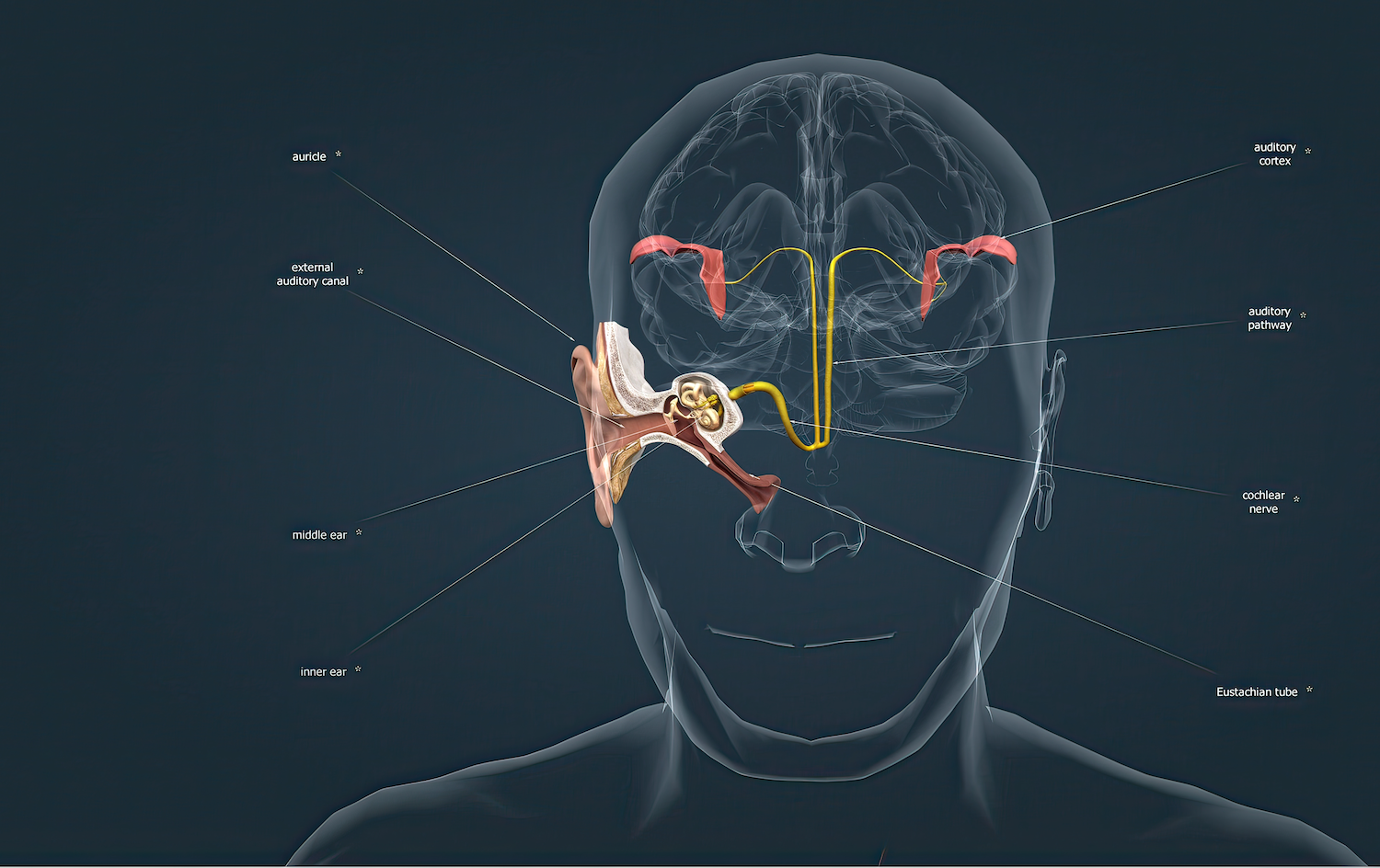
what is the eustachian tube and how does it work?
Click to enlarge.

The Eustachian tube is a tunnel connecting the back of the throat (the nasopharynx, specifically), to the space behind the eardrum (the middle ear). Essentially, the Eustachian tube’s function is to maintain an air pressure in the middle ear equal to that of the outside world and to allow fluid to drain from the middle ear space to the throat. In optimal conditions, when the air pressure in the middle ear equals that of the outside world, the tympanic membrane is in a neutral position and can transmit vibrations from sound waves efficiently to the bones of the middle ear and from there to the inner ear. The Eustachian tube orifice (entrance) is normally closed (collapsed) but normally opens briefly to allow air or fluid to pass. Air of the middle ear space is constantly being absorbed by the lining of the middle ear and mastoid, and consequently, air must periodically be replaced by the Eustachian tube temporarily opening so that air can pass from the throat to the middle ear space. An occasional popping sound of the ears, possibly accompanied by the feeling of a pressure change, is normal, healthy Eustachian tube function. When an air pocket moves in or out of the middle ear space rapidly, the position of the eardrum changes rapidly. Since the eardrum remains attached to the middle ear bones (ossicles), which are attached to the inner ear, the movement creates a signal to the brain interpreted as sound. The eardrum also has the sense of touch, so stretch and relaxation of the eardrum is associated with the sensory perception, adding to the feeling of an ear "popping.”


Endoscopic view of the right Eustachian tube orifice. The arrow indicates the orifice, in its usual closed configuration. The arch-shaped raised surface around the orifice demonstrates the torus tubarius, which is the cartilage of the Eustachian tube. When the surface tissue (called mucosa) becomes swollen, the orifice becomes less able to open periodically.
What is Eustachian tube dysfunction?
Eustachian tube dysfunction (ETD) is a disorder where the Eustachian tube fails to maintain middle ear air pressure equal to that of the outside world and to allow any accumulated middle ear fluid to drain to the throat. Most commonly, this is because the Eustachian tube does not open as much as needed. High air pressure within the middle ear space tends to resolve reliably, since the anatomy makes air escape from middle ear to throat occur more readily than air going from throat to middle ear. Thus, any persisting problem of unequalized air pressures is almost always less air pressure in the middle ear space than in the ear canal and outside world. This situation tends to cause fluid to collect in the middle ear and/or the adjacent mastoid cells, a condition called otitis media with effusion/serous otitis media. Occasionally, if the Eustachian tube is stuck open (called a patulous Eustachian tube), the problems are different, including sound waves from the throat transmitting up the Eustachian tube to the eardrum and yielding an unpleasant way of hearing one’s own voice (called autophonia) and one’s breathing.
Visualization of the eardrum (tympanic membrane)
Assessment of Eustachian tube dysfunction is aided by physical examination, which usually includes examination of the eardrum (tympanic membrane) as viewed from the ear canal. Retraction of the ear canal, as if it has been sucked inward, is a common indication of Eustachian tube dysfunction.

Examination of the ear with a microscope.

View of a normal right eardrum (tympanic membrane). The tympanic membrane is partially translucent, demonstrates normal blood vessels, color, and light reflectivity, Photo attribution to Michael Hawke MD.
What causes Eustachian tube dysfunction?
Both children and adults can develop Eustachian tube dysfunction. Children are especially predisposed to Eustachian tube dysfunction because their Eustachian tubes are narrow, more horizontal, and shorter than in adults.
Genetics also play a role in predisposing to Eustachian tube dysfunction. Some families’ genes for certain proteins present on the surface of the Eustachian tube are more favorable for bacteria to live and spread from the throat to the Eustachian tube and on up to the middle ear.
Infection of the throat may lead to swelling of throat structures including the Eustachian tube openings, making them less functional. Chronic infection of the adenoids frequently promotes Eustachian tube dysfunction in children.
Even among adults, whose Eustachian tubes are full grown and angled slightly downwards from ear to throat, various conditions causing narrowing of the Eustachian tube passageway lead to its dysfunction. Common causes of this include laryngopharyngeal reflux of stomach contents to the throat and Eustachian tube, surface irritants such as smoke, and throat infection or sinus infection.
Scar tissue of the Eustachian tube, which may develop from repeated infections or radiation therapy to the area, also cause inadequate opening of the Eustachian tube.
Uncommonly, a tumor (such as nasopharyngeal carcinoma) may block a Eustachian tube. Persistent one-sided Eustachian tube dysfunction can be a hint of possible anatomic obstruction of one Eustachian tube by tumor, prompting endoscopic visualization of the nasopharynx.
Most Eustachian tube dysfunction is related to inability of the Eustachian tube orifice (entrance) to open adequately. At the other end of the spectrum, where the Eustachian tube fails to work properly because it is open persistently—called patulous Eustachian tube—tends to occur in older individuals and/or people who have lost significant amounts of weight.
How is Eustachian tube dysfunction identified and what problems can it cause?
Symptoms alone may be adequate to identify Eustachian tube dysfunction. For example, most people will develop a sense of ear pressure or fullness and muffled hearing with a change in altitude, but if these symptoms fail to resolve within an hour or so at a given altitude, it would indicate at least mild Eustachian tube dysfunction. When the Eustachian tube fails to adjust air pressure of the middle ear to equalize the air pressure of the ear canal an outside world, the eardrum will naturally bulge outward or retract inward, usually causing mild hearing loss, until pressure on each side of the eardrum is equalized. If the pressure is not equalized, fluid may develop behind the eardrum (the middle ear space), causing moderate (but reversible) hearing loss. Uninfected fluid in the middle ear space is called serous otitis media. Frequent or long-standing development of fluid in the middle ear might go on to become a middle ear infection. A physician’s examination of the ear including visualization of the tympanic membrane may demonstrate these or other conditions caused by Eustachian tube dysfunction. A device called a tympanographer can also evaluate movement of the eardrum in response to different air pressures to produce a graph to aid in diagnosis. Lastly, various imaging studies such as CT or MRI may also demonstrate fluid in the middle ear or mastoid, or other conditions associated with Eustachian tube dysfunction (such as cholesteatoma) that would support a diagnosis of Eustachian tube dysfunction.
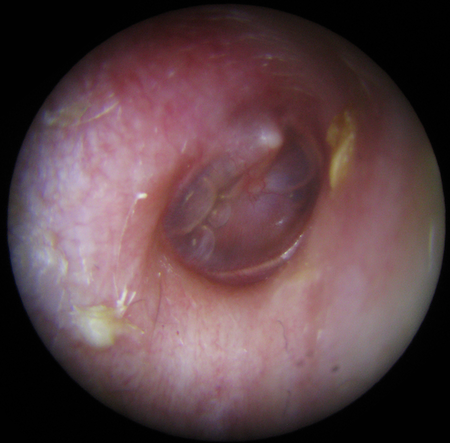
View of a right tympanic membrane with middle ear fluid (serous otitis media) seen through the tympanic membrane. This resulted from Eustachian tube dysfunction.

Severe retraction of the eardrum (tympanic membrane) as a result of Eustachian tube dysfunction.

View of a tympanic membrane with acute otitis media. Redness, thickening, bulging, and obscured landmarks of the eardrum (tympanic membrane) are evident. Photo attribution: B. Welleschik

how to get the most from your appointment for hearing
Appointment time is valuable. Here are some suggestions to make the most of your appointment. This preparation will help you and your doctor maximize efficiency and accuracy, freeing up time for questions and answers.
• Click here to prepare for your hearing loss appointment (adult)

This page